Logo Oapen
  • Search
  • Join
    • Deposit
    • For Librarians
    • For Publishers
    • For Researchers
    • Funders
    • Resources
    • OAPEN
    • For Librarians
    • For Publishers
    • For Researchers
    • Funders
    • Resources
    • OAPEN
    View Item 
    •   OAPEN Home
    • View Item
    •   OAPEN Home
    • View Item
    JavaScript is disabled for your browser. Some features of this site may not work without it.

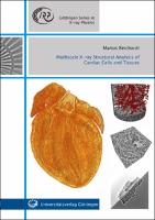
    Multiscale X-ray Structural Analysis of Cardiac Cells and Tissues

    Thumbnail
    Download PDF Viewer
    Book License
    Author(s)
    Reichardt, Marius
    Language
    English
    Show full item record
    Abstract
    The cardiac function relies on an intricate molecular and cellular three-dimensional (3d) architecture of a complex, dense and co-dependent cellular network. Structural alterations of the cardiac structure can affect its essential function and lead to severe dysfunction of the organ. Cardiovascular diseases are the main cause of death worldwide with a rising incidence. However, it is not possible to give a generalized answer how the heart is formed. Up to now, cardiac structure as well as physiologic and disease-related tissue alterations of the tissue are mainly investigated by established 2d imaging methods such as optical microscopy or electron microscopy. This work presents a multiscale and multimodal X-ray imaging approach, which allows to probe the heart structure from the scale of entire intact murine hearts to the molecular organisation of the sarcomer structure. While the molecular structure of the actomyosin complex is probed by scanning X-ray diffraction, the 3d arrangement of the cellular network is investigated by propagation-based X-ray phase-contrast tomography. In this context, the concept of 3d virtual histology of cardiac tissue by X-ray phase-contrast tomography using laboratory sources as well as highly coherent synchrotron radiation is being further developed.
    URI
    https://library.oapen.org/handle/20.500.12657/53509
    Keywords
    X-ray; cardiac cells; cardiac tissues
    ISBN
    978-3-86395-536-6
    Publisher
    Universitätsverlag Göttingen
    Publication date and place
    2022
    Classification
    Physics
    Rights
    http://creativecommons.org/licenses/by-sa/4.0/deed.de
    • Harvested from Göttingen

    Browse

    All of OAPENSubjectsPublishersLanguagesCollections

    My Account

    LoginRegister

    Export

    Repository metadata
    Logo Oapen
    • For Librarians
    • For Publishers
    • For Researchers
    • Funders
    • Resources
    • OAPEN

    Newsletter

    • Subscribe to our newsletter
    • view our news archive

    Follow us on

    Credits

    • logo Scoss
    • logo EU
    • logo Scoss
    • This project received funding from the European Union's Horizon 2020 research and innovation programme under grant agreement No 683680, 810640, 871069 and 964352.

    OAPEN is based in the Netherlands, with its registered office in the National Library in The Hague.

    Director: Niels Stern

    Address:
    OAPEN Foundation
    Prins Willem-Alexanderhof 5
    2595 BE The Hague
    Postal address:
    OAPEN Foundation
    P.O. Box 90407
    2509 LK The Hague

    Websites:
    OAPEN Home: www.oapen.org
    OAPEN Library: library.oapen.org
    DOAB: www.doabooks.org

    ©2020 OAPEN


    Export search results

    The export option will allow you to export the current search results of the entered query to a file. Differen formats are available for download. To export the items, click on the button corresponding with the preferred download format.

    A logged-in user can export up to 15000 items. If you're not logged in, you can export no more than 500 items.

    To select a subset of the search results, click "Selective Export" button and make a selection of the items you want to export. The amount of items that can be exported at once is similarly restricted as the full export.

    After making a selection, click one of the export format buttons. The amount of items that will be exported is indicated in the bubble next to export format.